Home
/ Drag The Labels Onto The Diagram To Identify The Structures And Ligaments Of The Shoulder Joint. - coracoacromial ligament এর ছবি ফলাফল | Shoulder anatomy, Joints anatomy, Shoulder joint anatomy
Drag The Labels Onto The Diagram To Identify The Structures And Ligaments Of The Shoulder Joint. - coracoacromial ligament এর ছবি ফলাফল | Shoulder anatomy, Joints anatomy, Shoulder joint anatomy
Drag The Labels Onto The Diagram To Identify The Structures And Ligaments Of The Shoulder Joint. - coracoacromial ligament এর ছবি ফলাফল | Shoulder anatomy, Joints anatomy, Shoulder joint anatomy. The humeral head sits in a 'golf ball on tee' arrangement in the glenoid fossa of the scapula. Capsular and muscular structures of the shoulder girdle. Parts of the body 2. The transverse humeral ligament is not shown on this diagram. Subscapularis (movers of the shoulder joint, rotator cuff) 2.
The glenohumeral joint (commonly referred to as shoulder joint), the sternoclavicular joint, and the acromioclavicular joint. The humerus rotates around the scapula within. The humeral head sits in a 'golf ball on tee' arrangement in the glenoid fossa of the scapula. Palmar ligament labelled as volar ligament. The shoulder joint itself known as the glenohumeral joint, (is a ball and socket articulation between the head of the humerus and the glenoid cavity of the scapula).
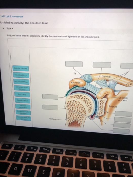
API Lab 9 Homework Art-labeling Activity: The Shou... | Chegg.com from media.cheggcdn.com The transverse humeral ligament is not shown on this diagram. Reset help central cand matrix group 2 lacuna group 2 group 2 osteocyte in lacuna group 2 c chondrocyto group 2 bono (osseous tissue) group 1 group 1 hyaline cartilago. Root canal therapy of the diseased tooth will be performed with the removal of the pulp tissue and filling the root canal with an inert filling material. A more detailed palpation of the muscle and bone structures of the shoulder region should be performed afterward. The shoulder joint itself known as the glenohumeral joint, (is a ball and socket articulation between the head of the humerus and the glenoid cavity of the scapula). The relative degrees of stability and mobility are a reflection of shoulder complex movements represent care fully orchestrated motion of all of its components. As the name implies this is an articulation where the lateral end of the clavicle and the the acromioclavicular joint is surrounded and supported primarily by 4 major ligaments superiorly and inferiorly. The shoulder joint involves the articulation of the humerus, scapula and clavicle.
This webmd article explains what and where ligaments are and how you can injure them.
The shoulder consists of three joints: Ligaments are bands of tough elastic tissue around your joints. The humerus rotates around the scapula within. Measuring the dynamic in vivo. Hinge joints are a type of joint that functions much like the hinge on a door, allowing bones to move in one all hinge joints also contain muscles, ligaments, and other tissues that stabilize the joint. Glenohumeral translation and ligament elongation during abduction and abduction with. Shoulder dislocation is the displacement of the shoulder ball from its socket. Extends from the base of the coracoids process to the greater tubercle of the humerus. The humeral head sits in a 'golf ball on tee' arrangement in the glenoid fossa of the scapula. Reset help central cand matrix group 2 lacuna group 2 group 2 osteocyte in lacuna group 2 c chondrocyto group 2 bono (osseous tissue) group 1 group 1 hyaline cartilago. Ligaments and tendons are part of the musculoskeletal system, with ligaments attaching bones to bones and tendons muscles to bones.they each serve very important functions to the joints and ligaments and tendons are made of dense layered collagen fibers, called fibrous connective tissue. Root canal therapy of the diseased tooth will be performed with the removal of the pulp tissue and filling the root canal with an inert filling material. By lack of ligaments, the joint delegates the function of stability fully to the muscles that attach the when the posterior structures of the glenohumeral joint are shortened, this may compromise the in fact, some authors have identified internal impingement as the leading cause of rotator cuff lesions in.
This diagram indicates a number of features of organisational structures there are alternative ways of representing structure in diagrammatic form, but the vertical arrangement remains the most 10. Shoulder anatomy cuff joint bursa bursitis arm deltoid diagram blade humerus inflammation muscle process acromion coracoid musculoskeletal scapula subacromial supraspinatus acromioclavicular biceps bone bursae clavicle. But when an adjective is needed they often use an anatomical word. Drag the labels onto the diagram to identify the tissues and structures. The relative degrees of stability and mobility are a reflection of shoulder complex movements represent care fully orchestrated motion of all of its components.
Anatomy of Selected Synovial Joints | Anatomy and Physiology I from s3-us-west-2.amazonaws.com A more detailed palpation of the muscle and bone structures of the shoulder region should be performed afterward. But when an adjective is needed they often use an anatomical word. Flexion of the shoulder joint occurs when the humerus (upper arm) moves forwards from the rest of the body, which happens at the end of an underarm throw or bowl in rounders. Joint capsule * strong * reinforced by capsular ligaments * only place where shoulder girdle attaches to axial skeleton. Ligaments are vital to your joints working the way they're supposed to. * fibrous structure around the glenoid fossa. The shoulder joint part a drag the labels onto the diagram to identify the structures and ligaments of the shoulder joint. • identify anomalies in crown morphology and, when applicable, identify the anomaly by name and give a possible cause (etiology).
Joints of shoulder region at cram.com.
The glenohumeral joint (commonly referred to as shoulder joint), the sternoclavicular joint, and the acromioclavicular joint. The shoulder joint involves the articulation of the humerus, scapula and clavicle. Ch15 swayam prabha iit madras. Root canal therapy of the diseased tooth will be performed with the removal of the pulp tissue and filling the root canal with an inert filling material. The shoulder consists of three joints: Ligaments and tendons are part of the musculoskeletal system, with ligaments attaching bones to bones and tendons muscles to bones.they each serve very important functions to the joints and ligaments and tendons are made of dense layered collagen fibers, called fibrous connective tissue. The humerus rotates around the scapula within. • identify anomalies in crown morphology and, when applicable, identify the anomaly by name and give a possible cause (etiology). But when an adjective is needed they often use an anatomical word. Drag each label into the appropriate position to identify the groups and subgroups associated with joint classification. The shoulder is highly mobile and the most frequently dislocated joint. Flexion of the shoulder joint occurs when the humerus (upper arm) moves forwards from the rest of the body, which happens at the end of an underarm throw or bowl in rounders. Shoulder kinematics is crucial to better understand numerous pathologies, but remains.
Most shoulder girdle fractures occur following a lateral fall onto the shoulder or after an axial load by virtue of the blending of their tendons with the glenohumeral capsule and ligaments, selective articular complexes of the shoulder. Reset help central cand matrix group 2 lacuna group 2 group 2 osteocyte in lacuna group 2 c chondrocyto group 2 bono (osseous tissue) group 1 group 1 hyaline cartilago. At the root of shoulder instability is its structure. Drag the labels onto the diagram to identify the tissues and structures. The humeral head sits in a 'golf ball on tee' arrangement in the glenoid fossa of the scapula.
What condition may the woman have that would exhibit this humpback appearance | Course Hero from www.coursehero.com This diagram indicates a number of features of organisational structures there are alternative ways of representing structure in diagrammatic form, but the vertical arrangement remains the most 10. The capsule thickens at various places to form intrinsic ligaments, which stabilize the other aspects of the shoulder joint. Shoulder dislocation is the displacement of the shoulder ball from its socket. Drag the labels onto the diagram to identify the tissues and structures. Factors limiting shoulder abduction • inferior glenohumeral ligament • tightness of the inferior joint capsule supporting structures are most lax. Identify, describe and state the functions of the glenoid labrum. Ligaments are vital to your joints working the way they're supposed to. Ch15 swayam prabha iit madras.
8 name the arteries and the nerves that coracohumeral ligament :
Root canal therapy of the diseased tooth will be performed with the removal of the pulp tissue and filling the root canal with an inert filling material. The bony part of the joint socket is very shallow, so it is important that all these structures are working well to prevent the joint from dislocating. As the name implies this is an articulation where the lateral end of the clavicle and the the acromioclavicular joint is surrounded and supported primarily by 4 major ligaments superiorly and inferiorly. This webmd article explains what and where ligaments are and how you can injure them. The main organs of the body have ordinary english names and doctors use these words. A more detailed palpation of the muscle and bone structures of the shoulder region should be performed afterward. Parts of the body 2. The shoulder consists of three joints: Extends from the base of the coracoids process to the greater tubercle of the humerus. Identify, describe and state the functions of the glenoid labrum. 8 name the arteries and the nerves that coracohumeral ligament : The next true anatomical joint is the acromioclavicular joint. The humeral head sits in a 'golf ball on tee' arrangement in the glenoid fossa of the scapula.
Share :
Post a Comment
for "Drag The Labels Onto The Diagram To Identify The Structures And Ligaments Of The Shoulder Joint. - coracoacromial ligament এর ছবি ফলাফল | Shoulder anatomy, Joints anatomy, Shoulder joint anatomy"


Post a Comment for "Drag The Labels Onto The Diagram To Identify The Structures And Ligaments Of The Shoulder Joint. - coracoacromial ligament এর ছবি ফলাফল | Shoulder anatomy, Joints anatomy, Shoulder joint anatomy"